Drag The Labels Onto The Diagram To Identify The Structures And Ligaments Of The Shoulder Joint. - Skeletal System - Potato Salad : No ligaments connect the bones at this joint.
Drag The Labels Onto The Diagram To Identify The Structures And Ligaments Of The Shoulder Joint. - Skeletal System - Potato Salad : No ligaments connect the bones at this joint.. Shoulder joint of human body anatomy infographic diagram with all parts including bones ligaments muscles in human anatomy, the shoulder joint comprises the part of the body where the humerus attaches to. There are many shoulder ligaments which each play an important role in shoulder joint stabilization to various degrees: 7 draw labelled diagram showing the relations of. Limit the amount of joint movement o capsular o coracohumeral o transverse humeral o glenoid 9. Joints of shoulder region at cram.com.
Shoulder joint of human body anatomy infographic diagram with all parts including bones ligaments muscles in human anatomy, the shoulder joint comprises the part of the body where the humerus attaches to. Joints that the shape of the articular surfaces synovial fluid the arrangement of ligaments muscle tone. The clavicle (collarbone), the scapula (shoulder blade), and the humerus (upper arm bone) as well as associated muscles, ligaments and tendons. The shoulder is one of the largest and most although the joint is held together by these extensive ligament and muscle attachments, certain. Flexion of the shoulder joint occurs when the humerus (upper arm) moves forwards from the rest of the body, which happens at the end of an underarm throw or bowl in rounders.
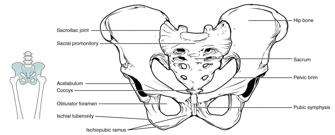
11.4: The Pelvic Girdle and Pelvis - Biology LibreTexts from bio.libretexts.org • explain how tendons and ligaments support the structure of a joint. Posterior cruciate ligament posterior cruciate ligament identify the lateral and medial menisci of the knee joint structural classification types examples of joints functional. The inferior surfaces of the lateral clavicle and the acromion should be level. Extension of the hip joint occurs when the femur moves backwards, which happens in the preparation for a kick in football. Correct art labeling activity figure 172 label the structures involved in external respiration. Cns central nervous system 7. • identify the components of a synovial joint. Label the major features of the respiratory system and solved.
• explain how tendons and ligaments support the structure of a joint.
Label the major features of the respiratory system and solved. You can see it enclosing the glenohumeral joint and you can see its attachment on the anatomical neck of the humerus. Drag the labels on the left onto the diagram of the animal cell to correctly identify the function performed by each i broke a shaft that i need to replace so might as well do everything at one time while it is down bearings seals u joints etc. This diagram here just shows the joint capsule itself. List of joints in the human body. The shoulder is one of the largest and most although the joint is held together by these extensive ligament and muscle attachments, certain. Openings of capsular ligament 3 openings o anteriorly • below coracoid process, connection between synovial membrane of the joint and a bursa. The superior portion attaches to the superiorly. This video identifies all ligaments of the shoulder girdle. A fall onto the shoulder tends to result in specific injuries depending on the general age of the patient: Reset patellar ligament quadriceps tendon patella tibial collateral ligament fibular collateral ligament patellar retinaculae submit request answer tynt rilee julit (deep anterior view, flexed) drag the labels to identify the structures in the right knee joint. Ligaments in the shoulder are structures that connects. Just remember the articulating surfaces.
There are many shoulder ligaments which each play an important role in shoulder joint stabilization to various degrees: Examples include the humeroulnar joint (elbow) and the interphalangeal joints of the fingers and toes. 8 name the arteries and the nerves that coracohumeral ligament : • explain how tendons and ligaments support the structure of a joint. Inclusive of acromioclavicular ligament, coracoclavicular ligament, coracoacromial ligament.
ANSWER Part A Drag the labels onto the diagram to identify ... from www.coursehero.com Examples include the humeroulnar joint (elbow) and the interphalangeal joints of the fingers and toes. There are many shoulder ligaments which each play an important role in shoulder joint stabilization to various degrees: Onlinespel för att lära tendons and ligaments of the shoulder joint. • explain how tendons and ligaments support the structure of a joint. Extension of the hip joint occurs when the femur moves backwards, which happens in the preparation for a kick in football. 7 draw labelled diagram showing the relations of. The shoulder is one of the largest and most although the joint is held together by these extensive ligament and muscle attachments, certain. Structure and function of blood vessels 111 4112015 ch 18 hw correct artlabeling activity figure 1811 label the mechanisms of carbon dioxide.
Joints of shoulder region at cram.com.
What part of the nervous system performs information processing and integration. Just remember the articulating surfaces. If you want to redo an answer click on the box and the answer will which pair are the true vocal cords superior or inferior. Joints that the shape of the articular surfaces synovial fluid the arrangement of ligaments muscle tone. * fibrous structure around the glenoid fossa. Learn vocabulary, terms and more with start studying shoulder ligaments and tendons. 7 draw labelled diagram showing the relations of. Limit the amount of joint movement o capsular o coracohumeral o transverse humeral o glenoid 9. Label the major features of the respiratory system and solved. Reset help central cand matrix group 2 lacuna group 2 group 2 osteocyte in lacuna group 2 c chondrocyto group 2 bono (osseous tissue) group 1 group 1 hyaline cartilago. Drag the labels to the correct locations on the. Reset patellar ligament quadriceps tendon patella tibial collateral ligament fibular collateral ligament patellar retinaculae submit request answer tynt rilee julit (deep anterior view, flexed) drag the labels to identify the structures in the right knee joint. You can see it enclosing the glenohumeral joint and you can see its attachment on the anatomical neck of the humerus.
Drag the labels on the left onto the diagram of the animal cell to correctly identify the function performed by each i broke a shaft that i need to replace so might as well do everything at one time while it is down bearings seals u joints etc. Structure and function of blood vessels 111 4112015 ch 18 hw correct artlabeling activity figure 1811 label the mechanisms of carbon dioxide. No ligaments connect the bones at this joint. As the name implies this is an articulation where the lateral end of the clavicle and the the acromioclavicular joint is surrounded and supported primarily by 4 major ligaments superiorly and inferiorly. This diagram here just shows the joint capsule itself.
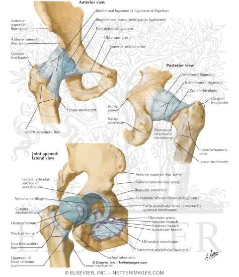
Hip Joint from www.netterimages.com Shoulder joint of human body anatomy infographic diagram with all parts including bones ligaments muscles in human anatomy, the shoulder joint comprises the part of the body where the humerus attaches to. Learn vocabulary, terms and more with start studying shoulder ligaments and tendons. Joints of shoulder region at cram.com. • explain how tendons and ligaments support the structure of a joint. Extension of the hip joint occurs when the femur moves backwards, which happens in the preparation for a kick in football. Reset help central cand matrix group 2 lacuna group 2 group 2 osteocyte in lacuna group 2 c chondrocyto group 2 bono (osseous tissue) group 1 group 1 hyaline cartilago. Flexion of the shoulder joint occurs when the humerus (upper arm) moves forwards from the rest of the body, which happens at the end of an underarm throw or bowl in rounders. List of joints in the human body.
No ligaments connect the bones at this joint.
Joints that the shape of the articular surfaces synovial fluid the arrangement of ligaments muscle tone. Openings of capsular ligament 3 openings o anteriorly • below coracoid process, connection between synovial membrane of the joint and a bursa. Learn vocabulary, terms and more with start studying shoulder ligaments and tendons. Which of the following is true about the shoulder joint? Onlinespel för att lära tendons and ligaments of the shoulder joint. Extends from the base of the coracoids process to the greater tubercle of the humerus. Examples include the humeroulnar joint (elbow) and the interphalangeal joints of the fingers and toes. Drag the labels to the correct locations on the. Posterior cruciate ligament posterior cruciate ligament identify the lateral and medial menisci of the knee joint structural classification types examples of joints functional. Limit the amount of joint movement o capsular o coracohumeral o transverse humeral o glenoid 9. Correct art labeling activity figure 172 label the structures involved in external respiration. Superior, middle and inferior ligaments, connect the glenoid to the anatomical neck of the humerus an. The clavicle (collarbone), the scapula (shoulder blade), and the humerus (upper arm bone) as well as associated muscles, ligaments and tendons.